Code Stroke
65 yo patient presented with sudden onset L arm weakness and confusion. MSU CT had revealed ischaemic CVA R MCA territory.
.HR Af 35-55, BP 100/60 (intermittently requiring metaraminol boluses. GCS 13-14 confused and agitated, unable to communicate a story. sats 100.
PMHx
Type B dissection repair 1 year ago, AF (on apixaban), HT, HC
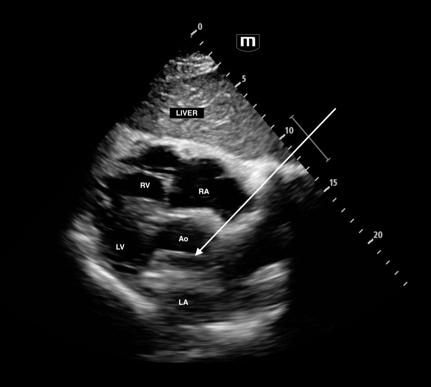
Given the hypotension and the hx of dissection, a bedside US was done and revealed:
subxyphoid 5 chamber view (ie transducer fanning anteriorly to reveal aorta)
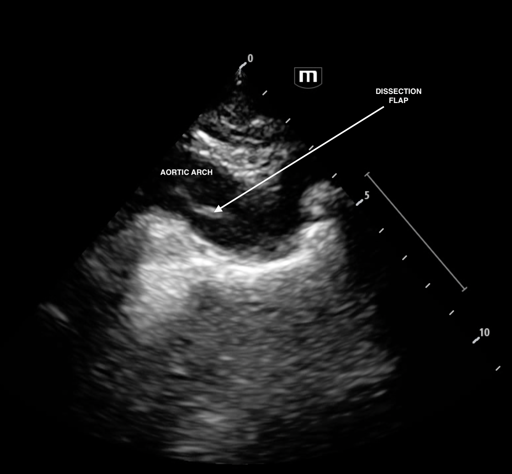
The dissection flap is seen more easily in the suprasternal view. The patient was fortunately NOT thrombolysed because of the bedside echo and went from CT to theatre in just under 2 hours and was successfully dc from hospital to rehab.
suprasternal view

Still of the subxyphoid from above with arrow pointing to dissection flap.

Still of the suprasternal cine loop from above with arrow pointing to dissection flap.
For more information on how to image type A dissection, click on the button below.
For info on how to get the suprasternal view click on the button below.
