Takatsubo
Takatsubo used to be a pretty rare diagnosis. However, its incidence seems to be increasing lately (1,2). It accounts for 2-3% of all acute patients presenting with acute coronary syndrome. Although it typically affects females >60yo (80-90%), 10% of cases occur in <50yo (predominantly males).
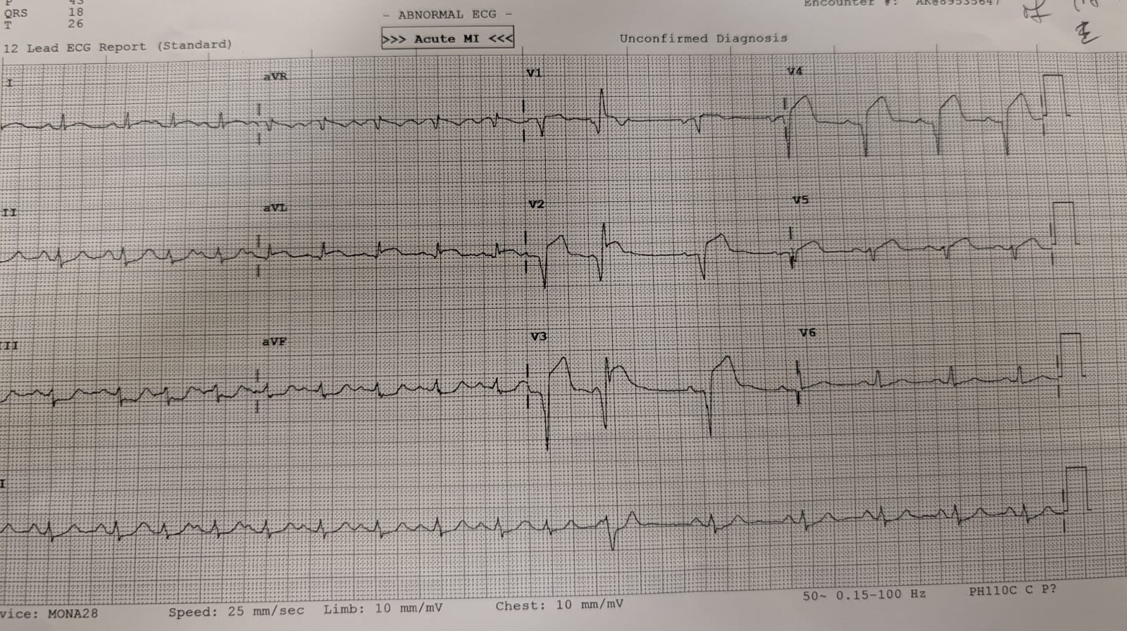
Takatsubo is left ventricular systolic failure due to anteroseptal and apical ballooning (1). Patients present with chest pain and sometimes features of pulmonary oedema, typically triggered by a stressful event. They may have an abnormal ECG with ST elevation in a non coronary distribution (3). At angiography, patients typically have normal coronaries; 20% may have evidence of vasospasm.

ECG of a patient with Takatsubo (courtesy of Ash Mukherjee
Although the LV dysfunction resolves completely, these patients can have an in-hospital mortality similar to STEMI patients (4).
The echo findings in takatsubo are classic:
1. Apical and anteroseptal hypokinesis and ballooning
2. Basal hyperkinesis - 20% of patients have evidence of dynamic outflow obstruction due to this.
Modified PLAx showing hyperkinetic basal segments and hypokinetic apex.
PSAx showing anterior and septal hypokinesis
Zoomed in view of LV from A4C
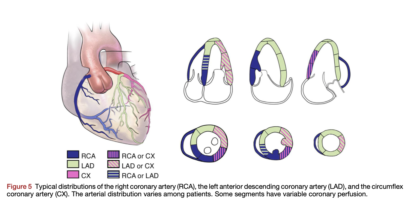
In Takatsubo, the RWMA typically doesn't follow a typical coronary distribution.

From Lang RM, Badano LP, Mor-Avi V, Afilalo J, Armstrong A, Ernande L, Flachskampf FA, Foster E, Goldstein SA, Kuznetsova T, Lancellotti P, Muraru D, Picard MH, Rietzschel ER, Rudski L, Spencer KT, Tsang W, Voigt JU. Recommendations for cardiac chamber quantification by echocardiography in adults: an update from the American Society of Echocardiography and the European Association of Cardiovascular Imaging. J Am Soc Echocardiogr. 2015 Jan;28(1):1-39
In RWMA due to ischaemia, the RWMA affects both the basal and apical segments of the wall. Ballooning is rare with acute ischaemia unless there is myocardial rupture and typically occurs 2-4 weeks post infarction with aneurysm formation. Comparitively, in Takatsubo, there is obvious ballooning and the distal (apical) part of the walls are hypokinetic and the basal segments of the same walls are hyperkinetic.
Nevertheless, given the high risk population, these patients are usually taken to the cath lab for further investigation. Further, despite normal coronary vasculature, they may have a moderate troponin rise.
REFERENCES
1. Singh T, Khan H, Gamble DT, Scally C, Newby DE, Dawson D. Takotsubo Syndrome: Pathophysiology, Emerging Concepts, and Clinical Implications. Circulation. 2022 Mar 29;145(13):1002-1019
2. Minhas AS, Hughey AB, Kolias TJ. Nationwide trends in reported incidence of takotsubo cardiomyopathy from 2006 to 2012. Am J Cardiol.
3. Namgung J. Electrocardiographic findings in takotsubo cardiomyopathy: ECG evolution and its difference from the ECG of acute coronary syndrome. Clin Med Insights Cardiol. 2014;8:29–34.
3. Templin C, Ghadri JR, Diekmann J, Napp LC, Bataiosu DR, Jaguszewski M, Cammann VL, Sarcon A, Geyer V, Neumann CA, et al. Clinical features and outcomes of takotsubo (stress) cardiomyopathy. N Engl J Med.
