Sudden onset vision loss
A young patient presented with sudden onset visual disturbance in her left eye. She felt like there was a film over her eye, as though straight lines were curved.
No hx of trauma or coagulopathy. She was myopic, unsure how severe.
OE
CN intact, PEARL no RAPD. Nil diplopia. L nasal superior quadrant VF defect L eye.
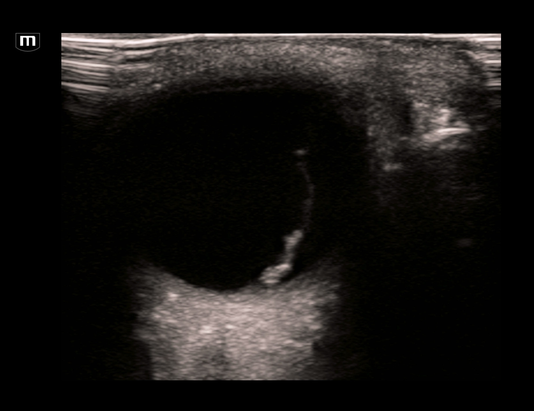
The EDUS showed this:

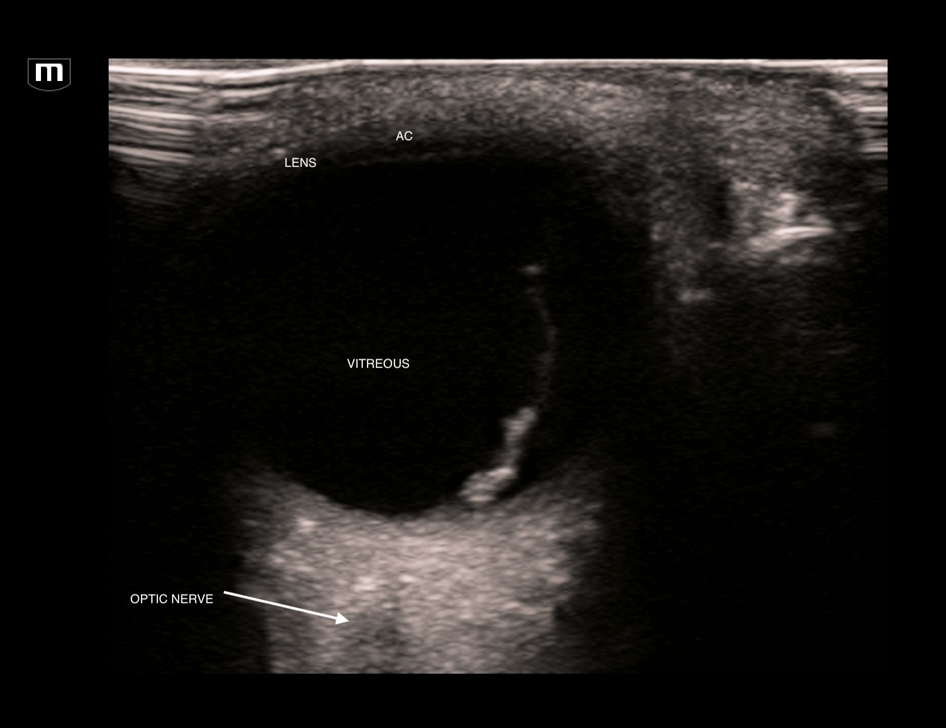
L orbit trans (annotated below)

This scan shows a thick hyperechoic membrane within the anechoic vitreous seeming to originate from the optic disc. This is typical of retinal detachment. Always fan through the orbit to try and image the optic nerve coming in to the orbit. A retnal detachment will be seen to be tethered to the optic disc. It does not cross over to the other side.
Clip below of imaging to define the optic nerve and the fact that the membrane does not cross the optic disc.
Clip of retinal detachment showing optic nerve and membrane tethered to the optic disc
Getting the patient to move their eye side to side will accentuate the detachment and highlight its mobility
undulating retinal detachment membrane
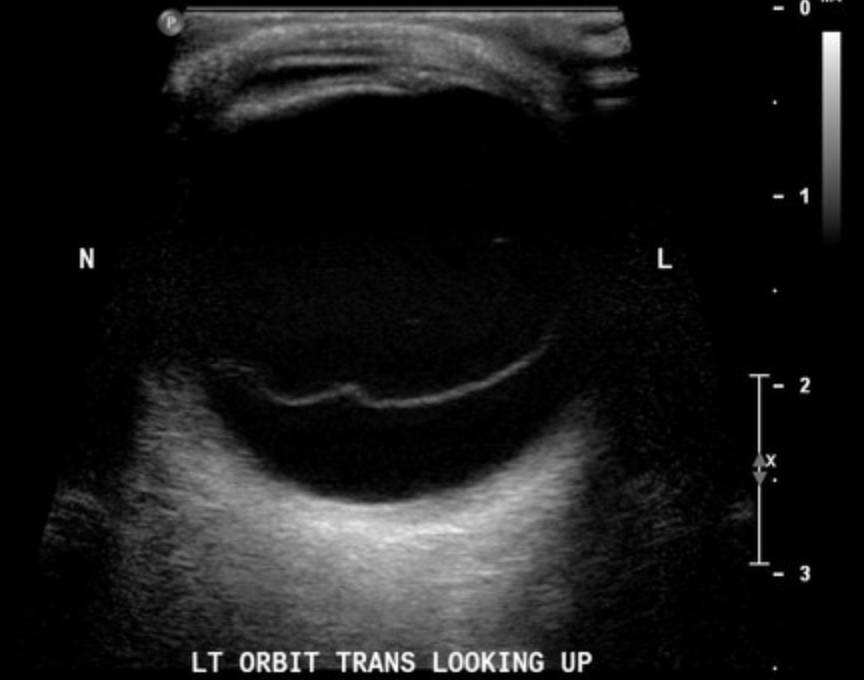
The main DDx for retinal detachment is posterior vitreous detachment (PVD). The US in PVD also shows a hyperechoic membrane in the vitreous. However, it is usually horizontal, thin and wispy and you need high gain to visualise it well. The membrane usually crosses anterior to the optic disc.

vitreous detachment (from radiopedia)
